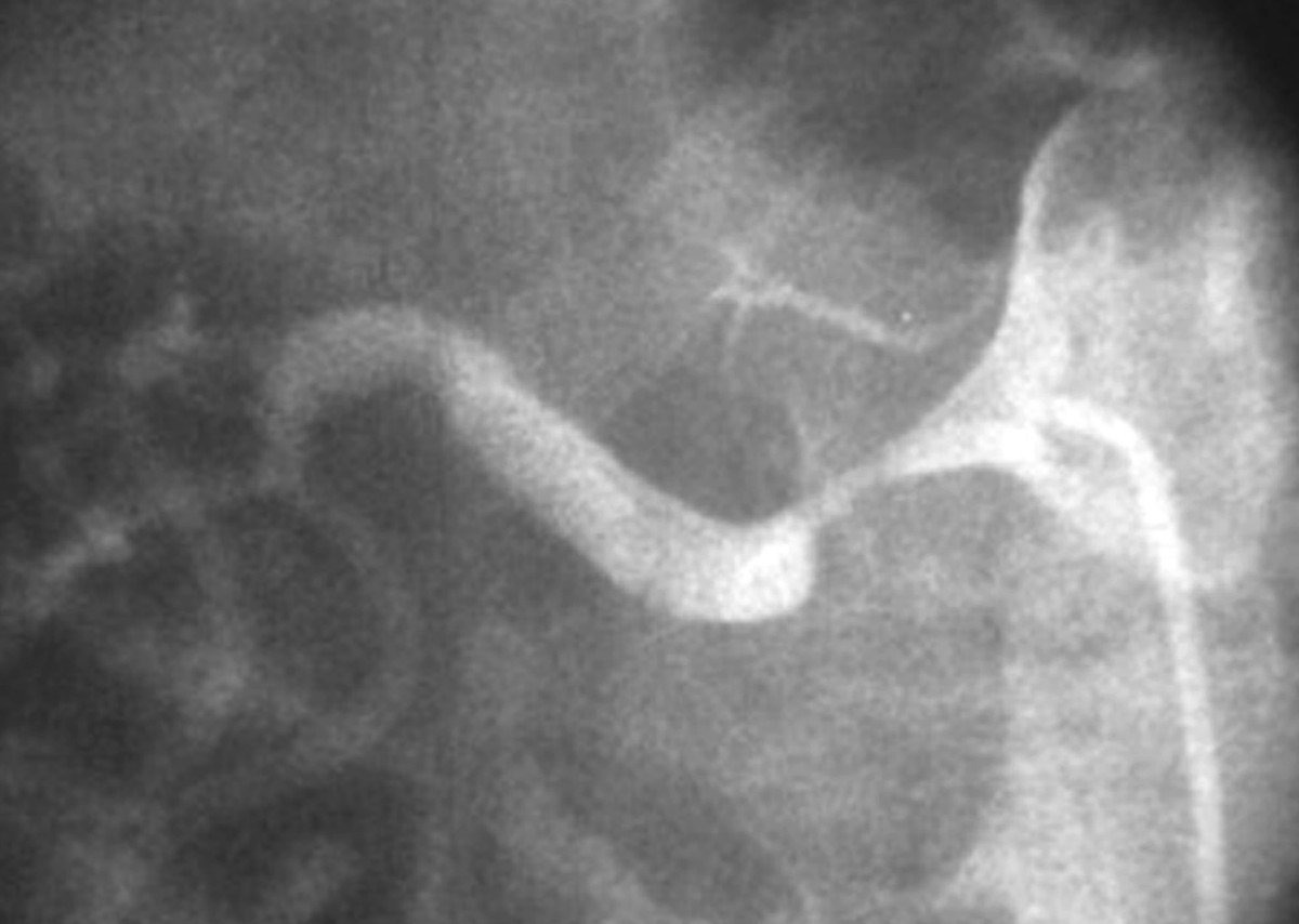
L'ecocolorDoppler è una procedura diagnostica non invasiva, ripetibile e relativamente economica che negli ultimi anni, in mani esperte, si è accreditata sempre più come ottimo strumento di screening di malattia nefrovascolare. Per tale motivo l'arteriografia riveste sempre più un ruolo interventistico e solo di rado diagnostico. Mentre la scintigrafia con test al captopril è stata utilizzata quasi esclusivamente nel passato, l'ecocolorDoppler delle arterie renali, l'angioTC e/o l'angioRM hanno sostituito le altre modalità di screening in molti centri. I test di screening per SAR sono migliorati considerevolmente durante l'ultimo decennio. Lo screening per SAR è indicato nel sospetto di ipertensione nefrovascolare o di nefropatia ischemica al fine di identificare i pazienti in cui è indicato un intervento di rivascolarizzazione. Per tale motivo, la diagnosi precoce di SAR è un obiettivo clinico importante poiché la terapia interventistica può migliorare o curare l'ipertensione e preservare la funzione renale. La SAR su base aterosclerotica è una malattia progressiva che può determinare in maniera asintomatica o paucisintomatica perdita graduale della funzione renale. Classicamente si presenta in una delle seguenti tre forme: stenosi dell'arteria renale (SAR) asintomatica, associata a ipertensione nefrovascolare e/o con nefropatia ischemica. SommarioLa malattia nefrovascolare è un disordine complesso e le cause più comuni sono la malattia aterosclerotica e la displasia fibromuscolare. However, when a discrepancy exists between clinical data and the results of Doppler US, additional tests are mandatory. Moreover, the evaluation of the resistive index (RI) at Doppler US may be very useful in RAS affected patients for predicting the response to revascularization.

Color-Doppler US (CDUS) is a noninvasive, repeatable, relatively inexpensive diagnostic procedure which can accurately screen for renovascular diseases if performed by an expert. An arteriogram is rarely needed for diagnostic purposes only. While captopril renography was widely used in the past, Doppler ultrasound (US) of the renal arteries (RAs), angio-CT, or magnetic resonance angiography (MRA) have replaced other modalities and they are now considered the screening tests of choice.

Screening tests for RAS have improved considerably over the last decade. Screening for RAS is indicated in suspected renovascular hypertension or ischemic nephropathy, in order to identify patients in whom an endoluminal or surgical revascularization is advisable. Thus, early diagnosis of RAS is an important clinical objective since interventional therapy may improve or cure hypertension and preserve renal function. Particularly, the atherosclerotic form is a progressive disease that may lead to gradual and silent loss of renal function. It can be found in one of three forms: asymptomatic renal artery stenosis (RAS), renovascular hypertension, and ischemic nephropathy. Low probability of renal artery stenosis (or, elevated renal artery to aortic ratio may represent renal artery stenosis recommend clinical correlation).Renovascular disease is a complex disorder, most commonly caused by fibromuscular dysplasia and atherosclerotic diseases. The pre-void bladder volume is _cc and the post-void residual volume is _cc (< 100 is normal).Ģ.

The bladder is normal in appearance and its wall measures _mm (< 3 is normal). The peak systolic velocity is _cm/sec and the renal artery to aortic ratio is _. It is normal in echotexture and demonstrate no evidence of hydronephrosis. The left kidney measures _cm in length with a cortical thickness of _cm. The peak systolic velocity is _m/sec (< 1.8 is normal) and the renal artery to aortic ratio is _ (< 3 is normal). It is normal in echotexture and demonstrates no evidence of hydronephrosis. The right kidney measures _cm in length (10-13 is normal in adults) with a cortical thickness of _cm (> 1 is normal). Multiple sonographic images of the kidneys and bladder were assessed for gray scale appearance and color doppler flow.


 0 kommentar(er)
